Determining whether a tooth requires a root canal rather than a simple filling involves evaluating specific clinical signs. Persistent, intensifying pain and heightened sensitivity to temperature changes often indicate deeper pulp involvement. Additional indicators such as localized swelling, gum tenderness, or discoloration suggest advanced infection or damage. Understanding these symptoms is critical for appropriate treatment selection, but several subtle factors must also be considered to avoid misdiagnosis or delayed intervention.
Persistent Tooth Pain That Worsens Over Time
Persistent tooth pain that intensifies over time is a primary indicator of underlying dental pulp inflammation or infection. This progressive pain often manifests as a dull ache or throbbing sensation localized to the affected tooth, distinguishing it from transient discomfort. Accompanying tooth sensitivity may initially respond to stimuli but escalates as the condition worsens. Effective pain management becomes increasingly challenging, as over-the-counter analgesics provide only temporary relief. The persistence and escalation of pain suggest that conservative treatments, such as fillings, are insufficient, indicating the need for endodontic intervention. Timely recognition of this symptom is critical to prevent further pulp necrosis and potential abscess formation. Consequently, monitoring the progression of tooth pain is essential for accurate diagnosis and determining the necessity of a root canal procedure.
Sensitivity to Hot and Cold Temperatures
In addition to escalating tooth pain, increased sensitivity to temperature extremes often signals compromised dental pulp health. When exposed to hot or cold stimuli, affected teeth may exhibit exaggerated temperature reactions due to inflamed or infected dental nerves within the pulp chamber. Unlike typical mild sensitivity, this heightened response can persist long after the stimulus is removed, indicating underlying nerve irritation or damage. Such sustained sensitivity suggests that the protective dentin and enamel layers are insufficient, allowing thermal stimuli to directly impact the pulp tissue. This condition frequently necessitates intervention beyond restorative fillings, as the pulp’s vitality is at stake. Accurate assessment of temperature reactions is critical for differentiating reversible pulpitis from irreversible damage requiring root canal therapy to prevent further complications.
Swelling and Tenderness Around the Tooth
Swelling of the gums surrounding a tooth often indicates an inflammatory response to infection or abscess formation. Tenderness upon palpation or biting pressure can signal compromised dental pulp or periapical tissue involvement. Accurate identification of these symptoms is critical for diagnosing the need for endodontic intervention.
Causes of Gum Swelling
Gum swelling and tenderness around a tooth often indicate an underlying dental issue that requires prompt evaluation. Common causes include localized infections stemming from dental caries that have progressed to the pulp, leading to periapical abscess formation. Inadequate oral hygiene can exacerbate plaque accumulation, contributing to gum disease, which manifests as inflammation and swelling. Periodontal pathogens induce an immune response, resulting in erythema and edema of the gingival tissues. Additionally, trauma or occlusal stress may provoke localized inflammatory reactions. Identifying these etiologies is critical, as untreated infections can compromise periodontal support and necessitate endodontic intervention such as a root canal. Hence, thorough clinical examination and radiographic assessment are essential to differentiate between reversible gingivitis and more severe pulpal or periapical pathology causing gum swelling.
Identifying Tooth Tenderness
Tenderness around a tooth often accompanies localized swelling and may indicate inflammation or infection within the pulp or surrounding periodontal structures. This symptom is a crucial diagnostic marker during a dental examination, as it often correlates with underlying pulpitis or apical periodontitis requiring prompt intervention. Tooth sensitivity to pressure or percussion, combined with tenderness, suggests compromised structural integrity or infection extending beyond superficial enamel damage. Accurate identification of these signs helps differentiate cases necessitating root canal therapy from those manageable by simple fillings. Clinicians rely on eliciting tenderness responses alongside radiographic and clinical findings to assess pulp health and periapical status. Early recognition of swelling and tenderness guarantees timely treatment, preventing progression to abscess formation or systemic involvement.
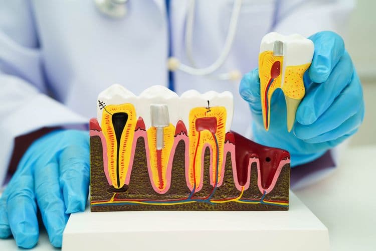
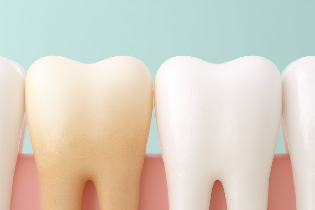
Discoloration or Darkening of the Affected Tooth
Discoloration or darkening of a tooth can result from intrinsic factors such as pulp necrosis or extrinsic staining due to dietary and environmental influences. Differentiating superficial stains from discoloration caused by underlying decay is essential for accurate diagnosis. Persistent darkening often indicates pulpal infection, which may necessitate root canal treatment.
Causes of Tooth Discoloration
Although tooth darkening can result from various factors, the primary cause in cases requiring a root canal is often internal pulp necrosis. This condition occurs when the dental pulp, containing nerves and blood vessels, undergoes irreversible damage or infection, leading to the breakdown of hemoglobin and subsequent deposition of dark pigments within the dentin. External factors such as consumption of staining substances or trauma may contribute but are less indicative of pulp health. Attempts at tooth whitening or natural remedies are typically ineffective against discoloration caused by internal necrosis, as these methods cannot penetrate to the source of pigment within the tooth structure. Accurate diagnosis is essential to determine whether discoloration necessitates endodontic intervention rather than superficial cosmetic treatments.
Differentiating Stains From Decay
Distinguishing between extrinsic stains and intrinsic decay is fundamental for accurate diagnosis and appropriate treatment planning. Stain types include superficial discolorations caused by dietary factors, tobacco, or poor oral hygiene, typically presenting as uniform and removable deposits on the tooth surface. In contrast, decay signs manifest as localized darkening or discoloration within the enamel or dentin, often accompanied by structural compromise such as cavitation or enamel breakdown. Unlike extrinsic stains, intrinsic discoloration from decay is not removable by polishing and indicates demineralization or bacterial invasion. Proper differentiation requires clinical examination supplemented by diagnostic tools like radiographs and transillumination to assess subsurface changes. Accurate identification of decay signs versus stain types guarantees timely intervention, preventing progression that may necessitate root canal therapy rather than a simple filling.
When Darkness Signals Infection
When a tooth exhibits a persistent darkening that extends beyond surface stains, it often indicates underlying infection or necrosis within the pulp tissue. Such darkness indicators are critical infection signs, suggesting compromised vascular supply and tissue death, which cannot be resolved by superficial treatments like fillings. Discoloration typically manifests as gray, brown, or black hues, reflecting internal hemorrhage or bacterial infiltration. This intrinsic staining is distinct from extrinsic stains caused by diet or hygiene. Clinicians assess these darkness indicators alongside symptoms such as sensitivity and pain to determine the necessity for root canal therapy. Early identification of these infection signs enables timely intervention, preventing further structural degradation and systemic complications. Consequently, persistent tooth discoloration serves as a significant diagnostic marker for pulp pathology requiring endodontic treatment.
Presence of a Pimple-Like Bump on the Gums
A pimple-like bump on the gums often indicates the presence of a dental abscess, which results from an infection within the tooth pulp or surrounding periodontal tissues. This localized swelling, also known as a parulis or gum boil, serves as a drainage point for pus accumulating due to bacterial invasion. Its appearance signals compromised gum health and the failure of the body’s immune response to contain the infection internally. Without appropriate intervention, such as a root canal treatment, the infection can progress, causing further destruction of the pulp and supporting structures. Identification of this symptom is critical, as it differentiates cases requiring endodontic therapy from those manageable by simple fillings, emphasizing the importance of prompt dental evaluation to prevent systemic complications and preserve oral integrity.
Pain When Chewing or Biting Down
The presence of a pimple-like bump on the gums often coincides with other indicators of dental pulp infection, such as discomfort during mastication. Pain when chewing or biting down is a significant clinical sign that distinguishes the need for root canal therapy from a simple filling. This symptom arises due to inflammation or infection within the tooth’s pulp chamber, causing heightened sensitivity to occlusal forces. Patients frequently report chewing discomfort and biting sensitivity localized to the affected tooth, which may intensify with pressure application. Unlike superficial enamel issues, these symptoms suggest deeper pulpal involvement, necessitating endodontic intervention to alleviate pain and prevent further tissue damage. Timely diagnosis based on these functional symptoms is essential to preserve tooth integrity and avoid complications.
Deep Decay Reaching the Tooth’s Pulp
Although dental decay often initiates in the enamel, progression into the dentin and eventual penetration of the pulp chamber signifies critical pathology requiring root canal treatment. Deep decay reaching the tooth’s pulp results in direct exposure of the pulp tissue to bacterial infiltration, causing pulp damage. This exposure frequently leads to inflammation, necrosis, and subsequent tooth infection. Unlike superficial carious lesions amenable to fillings, involvement of the pulp necessitates removal of infected tissue to prevent abscess formation and systemic complications. Clinical assessment, supplemented by radiographic imaging, confirms the extent of decay and pulp involvement. Timely intervention with root canal therapy eradicates infection, preserves tooth structure, and alleviates symptoms. Failure to address deep pulp involvement can result in persistent infection, tooth loss, and spread of infection beyond the local site.
Prolonged Pain After Dental Procedures or Trauma
Several cases of prolonged pain following dental procedures or trauma indicate possible pulp or periapical tissue damage requiring root canal therapy. Post procedure discomfort that persists beyond the expected healing period often signifies irreversible pulpitis or necrosis. Dental trauma, including fractures or luxations, can compromise the pulp’s vascular supply, leading to inflammation or infection. Such conditions manifest as persistent, throbbing pain, heightened sensitivity to thermal stimuli, or spontaneous discomfort unrelieved by analgesics. Radiographic examination may reveal periapical radiolucencies indicative of apical periodontitis. In these scenarios, conservative treatments like fillings prove insufficient, necessitating endodontic intervention to remove necrotic tissue and prevent further complications. Accurate diagnosis relies on clinical assessment combined with diagnostic tests to distinguish between reversible irritation and pulp necrosis requiring root canal therapy.
Frequently Asked Questions
How Long Does a Root Canal Procedure Take?
The root canal duration typically ranges from 60 to 90 minutes, depending on procedure steps including anesthetic administration, pulp removal, canal cleaning, shaping, and filling. Complex cases may require multiple visits, extending total treatment time.
Is a Root Canal More Expensive Than a Filling?
A root canal is typically more expensive than a filling due to its complexity and duration. Cost comparison should consider insurance coverage specifics, as policies often cover fillings more extensively, affecting out-of-pocket expenses for patients.
Can a Root Canal Be Done in One Visit?
A root canal can be completed in a single visit depending on the case complexity and infection severity. Treatment duration varies, but advancements in endodontic techniques often enable efficient, one-session procedures without compromising outcomes.
What Are the Risks of Not Getting a Root Canal?
Failure to undergo a root canal can result in worsening tooth infection, leading to abscess formation, systemic spread, and severe pain. Delayed treatment complicates pain management and may necessitate tooth extraction or extensive surgical intervention.
How Long Is the Recovery After a Root Canal?
Recovery after a root canal typically spans 3 to 7 days. Effective pain management includes prescribed analgesics, cold compresses, and avoiding hard foods. Recovery tips emphasize oral hygiene and follow-up visits to guarantee proper healing.